Compare brain regions
Choose an electrophysiology feature, find a brain region, and compare its values with other regions on the atlas.
 Ephys Atlas
International Brain Laboratory
Browse electrical features across brain regions and inspect their distributions alongside the anatomy.
The atlas runs in your browser without an account.
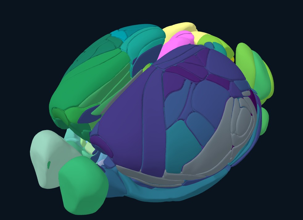
Anatomical views
Coronal, sagittal, and horizontal slices follow the same position in the brain.
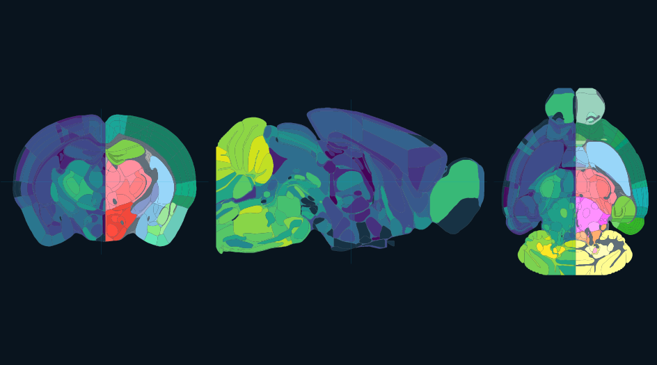
Choose an electrophysiology feature, find a brain region, and compare its values with other regions on the atlas.
View distributions and linked volume slices. Each dataset includes its source and release information.
Import a compatible dataset to use these tools with your own data. Local imports stay in your browser.
About the project
The IBL Ephys Atlas displays regional electrophysiology summaries, voxel-scale features, and brain-wide analysis results on the mouse brain atlas.
Developed by the International Brain Laboratory.
For dataset sources, release information, and provenance, open Data details in the viewer. Use these records when citing the data.
Open atlasThe anatomical reference comes from the Allen Mouse Brain Common Coordinate Framework (CCFv3), provided by the Allen Institute.
IBL receives support from Simons Foundation International, Simons Foundation, Wellcome, and the National Institutes of Health.
Loading atlas…